Click image to see more details
Product Info Summary
| SKU: | A00390-2 |
|---|---|
| Size: | 100 μg/vial |
| Reactive Species: | Human |
| Host: | Rabbit |
| Application: | ELISA, Flow Cytometry, IHC |
Customers Who Bought This Also Bought


Product info
Product Name
Anti-CCR7 Antibody Picoband™
SKU/Catalog Number
A00390-2
Size
100 μg/vial
Form
Lyophilized
Description
Boster Bio Anti-CCR7 Antibody Picoband™ catalog # A00390-2. Tested in ELISA, Flow Cytometry, IHC applications. This antibody reacts with Human.
Storage & Handling
At -20°C for one year from date of receipt. After reconstitution, at 4°C for one month. It can also be aliquotted and stored frozen at -20°C for six months. Avoid repeated freezing and thawing.
Cite This Product
Anti-CCR7 Antibody Picoband™ (Boster Biological Technology, Pleasanton CA, USA, Catalog # A00390-2)
Host
Rabbit
Contents
Each vial contains 4 mg Trehalose, 0.9 mg NaCl, 0.2 mg Na2HPO4.
Clonality
Polyclonal
Isotype
Rabbit IgG
Immunogen
E.coli-derived human CCR7 recombinant protein (Position: K332-E371).
*Blocking peptide can be purchased. Costs vary based on immunogen length. Contact us for pricing.
Cross-reactivity
No cross-reactivity with other proteins.
Reactive Species
A00390-2 is reactive to CCR7 in Human
Applications
A00390-2 is guaranteed for ELISA, Flow Cytometry, IHC Boster Guarantee
Observed Molecular Weight
38 kDa
Calculated molecular weight
42.874kDa
Background of CCR7
C-C chemokine receptor type 7 is a protein that in humans is encoded by the CCR7 gene. The protein encoded by this gene is a member of the G protein-coupled receptor family. This receptor was identified as a gene induced by the Epstein-Barr virus (EBV), and is thought to be a mediator of EBV effects on B lymphocytes. This receptor is expressed in various lymphoid tissues and activates B and T lymphocytes. It has been shown to control the migration of memory T cells to inflamed tissues, as well as stimulate dendritic cell maturation. The chemokine (C-C motif) ligand 19 (CCL19/ECL) has been reported to be a specific ligand of this receptor. Signals mediated by this receptor regulate T cell homeostasis in lymph nodes, and may also function in the activation and polarization of T cells, and in chronic inflammation pathogenesis. Alternative splicing of this gene results in multiple transcript variants.
Antibody Validation
Boster validates all antibodies on WB, IHC, ICC, Immunofluorescence, and ELISA with known positive control and negative samples to ensure specificity and high affinity, including thorough antibody incubations.
Innovating Scientists Reward
If you are the first to review this product, or if you have results for a special sample, species or application this product is not validated in, share your results with us and receive product credits you can use towards any Boster products! Applicable to all scientists worldwide.
Submit A Review
Assay dilution & Images
Reconsitution
Adding 0.2 ml of distilled water will yield a concentration of 500 μg/ml.
Assay Dilutions Recommendation
The recommendations below provide a starting point for assay optimization. The actual working concentration varies and should be decided by the user.
Immunohistochemistry(Paraffin-embedded Section), 2-5 μg/ml, Human
Flow Cytometry, 1-3 μg/1x106 cells, Human
Direct ELISA, 0.1-0.5 μg/ml, Human
Validation Images & Assay Conditions
Click image to see more details
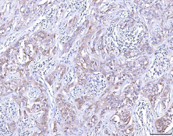
Figure 1. IHC analysis of CCR7 using anti-CCR7 antibody (A00390-2).
CCR7 was detected in a paraffin-embedded section of human lymphoma tissue. Heat mediated antigen retrieval was performed in EDTA buffer (pH 8.0, epitope retrieval solution). The tissue section was blocked with 10% goat serum. The tissue section was then incubated with 2 μg/ml rabbit anti-CCR7 Antibody (A00390-2) overnight at 4°C. Peroxidase Conjugated Goat Anti-rabbit IgG was used as secondary antibody and incubated for 30 minutes at 37°C. The tissue section was developed using HRP Conjugated Rabbit IgG Super Vision Assay Kit (Catalog # SV0002) with DAB as the chromogen.

Click image to see more details
Figure 2. Flow Cytometry analysis of U20S cells using anti-CCR7 antibody (A00390-2).
Overlay histogram showing U20S cells stained with A00390-2 (Blue line). The cells were blocked with 10% normal goat serum. And then incubated with rabbit anti-CCR7 Antibody (A00390-2, 1 μg/1x106 cells) for 30 min at 20°C. DyLight®488 conjugated goat anti-rabbit IgG (BA1127, 5-10 μg/1x106 cells) was used as secondary antibody for 30 minutes at 20°C. Isotype control antibody (Green line) was rabbit IgG (1 μg/1x106) used under the same conditions. Unlabelled sample (Red line) was also used as a control.
Protein Target Info & Infographic
Gene/Protein Information For CCR7 (Source: Uniprot.org, NCBI)
Gene Name
CCR7
Full Name
C-C chemokine receptor type 7
Weight
42.874kDa
Superfamily
G-protein coupled receptor 1 family
Alternative Names
BLR2; BLR2C-C chemokine receptor type 7; C-C CKR-7; CC-CKR-7; CCR7; CCR-7; CD197 antigen; CD197; CDw197; CDw197CC chemokine receptor 7; chemokine (C-C motif) receptor 7; CMKBR7; CMKBR7chemokine (C-C) receptor 7; EBI1; EBI1EBV-induced G protein-coupled receptor 1; EBV-induced G-protein coupled receptor 1; Epstein-Barr virus induced gene 1; Epstein-Barr virus induced G-protein coupled receptor; Epstein-Barr virus-induced G-protein coupled receptor 1; EVI1; lymphocyte-specific G protein-coupled peptide receptor; MIP-3 beta receptor CCR7 BLR2, CC-CKR-7, CCR-7, CD197, CDw197, CMKBR7, EBI1 C-C motif chemokine receptor 7 C-C chemokine receptor type 7|Bukitts lymphoma receptor 2|CC chemokine receptor 7|EBV-induced G protein-coupled receptor 1|Epstein-Barr virus induced gene 1|Epstein-Barr virus-induced G-protein coupled receptor 1|MIP-3 beta receptor|chemokine (C-C motif) receptor 7|lymphocyte-specific G protein-coupled peptide receptor
*If product is indicated to react with multiple species, protein info is based on the gene entry specified above in "Species".For more info on CCR7, check out the CCR7 Infographic

We have 30,000+ of these available, one for each gene! Check them out.
In this infographic, you will see the following information for CCR7: database IDs, superfamily, protein function, synonyms, molecular weight, chromosomal locations, tissues of expression, subcellular locations, post-translational modifications, and related diseases, research areas & pathways. If you want to see more information included, or would like to contribute to it and be acknowledged, please contact [email protected].
Specific Publications For Anti-CCR7 Antibody Picoband™ (A00390-2)
Hello CJ!
No publications found for A00390-2
*Do you have publications using this product? Share with us and receive a reward. Ask us for more details.
Recommended Resources
Here are featured tools and databases that you might find useful.
- Boster's Pathways Library
- Protein Databases
- Bioscience Research Protocol Resources
- Data Processing & Analysis Software
- Photo Editing Software
- Scientific Literature Resources
- Research Paper Management Tools
- Molecular Biology Software
- Primer Design Tools
- Bioinformatics Tools
- Phylogenetic Tree Analysis
Customer Reviews
Have you used Anti-CCR7 Antibody Picoband™?
Submit a review and receive an Amazon gift card.
- $30 for a review with an image
Be the first to review Anti-CCR7 Antibody Picoband™
*The first user to submit a review for a product is eligible for Boster's Innovating Scientists Reward, which gives product credits. This is in addition to the gift card reward.
Customer Q&As
Have a question?
Find answers in Q&As, reviews.
Can't find your answer?
Submit your question
